หัวข้อ: สิ่งที่เห็นในฟิล์ม X-ray อาจไม่ใช่ "ต้นเหตุที่แท้จริง" ของอาการปวดหลังเสมอไป ( English Ver. below)
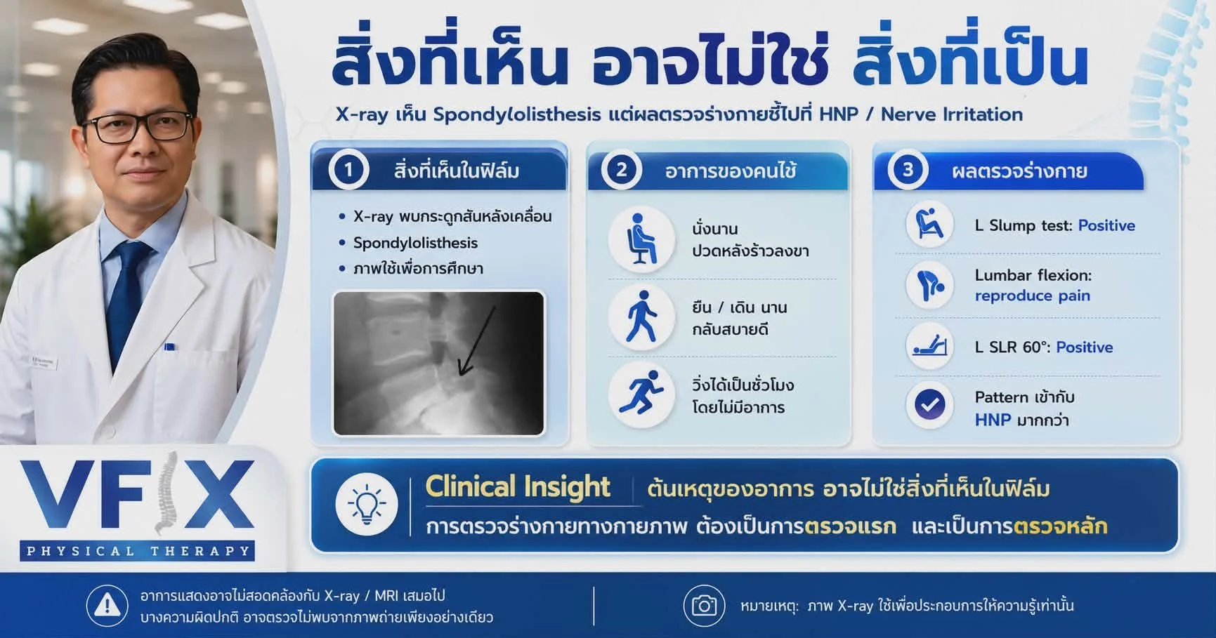
สิ่งที่เห็นอาจจะไม่ใช่สิ่งที่เป็น
คุณเคยสงสัยไหมครับว่า ทำไมอาการปวดหลัง ชา หรือปวดร้าวลงขาของคุณ ถึงไม่สอดคล้องกับผลฟิล์ม X-ray หรือ MRI ที่ออกมา?
วันนี้ผมมีกรณีศึกษาที่น่าสนใจจากคลินิกมาเล่าให้ฟังครับ เป็นเคสที่ยืนยันคำกล่าวที่ว่า "สิ่งที่เห็น อาจไม่ใช่สิ่งที่เป็นเสมอไป"
ความขัดแย้งระหว่าง "ภาพที่เห็น" กับ "อาการที่เป็น" เมื่อไม่นานมานี้ มีคนไข้เข้ามาปรึกษาพร้อมกับฟิล์ม X-ray กระดูกสันหลังส่วนเอว ภาพจากฟิล์มแสดงให้เห็นอย่างชัดเจนว่าคนไข้มีภาวะ กระดูกสันหลังเคลื่อน (Spondylolisthesis) หากดูแค่ภาพ เราอาจด่วนสรุปแนวทางการรักษาไปแล้ว แต่เมื่อทำการซักประวัติและตรวจร่างกายทางกายภาพเชิงลึก (Clinical Assessment) ความจริงอีกด้านก็ปรากฏขึ้น:
อาการของคนไข้: เวลานั่งนานๆ จะมีอาการปวดหลังร้าวลงขามากขึ้น แต่หากยืน เดิน หรือแม้แต่วิ่งต่อเนื่องเป็นชั่วโมง กลับรู้สึกสบายดีและไม่มีอาการปวดเลย
ผลการตรวจร่างกายทางคลินิก (Physical Examination):
Left Slump Test: Positive (ให้ผลบวก)
Lumbar Flexion: กระตุ้นให้เกิดอาการปวดร้าวลงสะโพกซ้าย
Left SLR (Straight Leg Raise) ที่ 60 องศา: Positive (ให้ผลบวก)
การวิเคราะห์ทางคลินิก (Clinical Reasoning) ผลการตรวจร่างกายทั้งหมดที่กล่าวมา ชี้ชัดไปที่ ภาวะหมอนรองกระดูกปลิ้นทับเส้นประสาท (HNP - Herniated Nucleus Pulposus) ไม่ใช่อาการที่เกิดจากกระดูกสันหลังเคลื่อนตามที่เห็นในฟิล์ม X-ray (ซึ่งหากอาการหลักมาจากกระดูกสันหลังเคลื่อน ลักษณะอาการแสดงจะเป็นอีกรูปแบบหนึ่ง)
ทำไมการตรวจร่างกายจึงสำคัญที่สุด? นี่คือเหตุผลที่ในการทำกายภาพบำบัดเฉพาะทาง เรายึดหลักการที่ว่า "การตรวจร่างกายทางกายภาพ ต้องเป็นการตรวจแรกและเป็นการตรวจหลักเสมอ" ภาพถ่ายทางรังสี (X-ray/MRI) เป็นเครื่องมือประกอบการวินิจฉัยที่ยอดเยี่ยม แต่ไม่สามารถแทนที่การตรวจร่างกายได้ เพราะ:
อาการเจ็บปวดของคนไข้ อาจไม่สอดคล้องกับความผิดปกติที่เห็นใน MRI หรือ X-ray เสมอไป
ความผิดปกติบางอย่างที่ทำให้เกิดการระคายเคืองของเส้นประสาท อาจตรวจไม่พบจากการถ่ายภาพเพียงอย่างเดียว
ในโลกของการวินิจฉัยโรค สิ่งที่เห็นในแผ่นฟิล์มอาจเป็นเพียงผลพลอยได้ (Incidental finding) แต่ร่องรอยที่แท้จริงของความเจ็บปวด จะถูกเปิดเผยผ่านการประเมินการเคลื่อนไหวและการตรวจร่างกายอย่างละเอียดเท่านั้น
การรักษาที่แม่นยำและตรงจุด จึงต้องเริ่มต้นจากการตรวจแยกอาการอย่างถูกต้องครับ

Symptoms do not always correlate with X-ray / MRI findings
Title: What You See on an X-Ray Isn't Always the Root Cause of Back Pain
Have you ever experienced lower back pain, numbness, or sciatica, only to find that your X-ray or MRI results don’t quite match the symptoms you're feeling?
There is a profound truth in specialized physical therapy: What you see on the film is not always the root cause of the problem. A recent clinical case at our practice perfectly illustrates this point.
The Disconnect Between Imaging and Symptoms A patient recently consulted us, bringing along a lumbar spine X-ray. The imaging clearly showed Spondylolisthesis (a slipped vertebra).
If we had relied solely on the X-ray, the treatment plan would have been vastly different. However, an in-depth clinical interview and physical examination revealed a completely different story:
The Patient's Experience: Sitting for prolonged periods exacerbated the back pain, radiating down the leg. Paradoxically, standing, walking, or even running for an hour felt perfectly fine with zero discomfort.
The Clinical Findings:
Left Slump Test: Positive
Lumbar Flexion: Reproduced pain radiating down to the left buttock
Left SLR (Straight Leg Raise) at 60 degrees: Positive
The Clinical Reasoning These specific signs and physical responses are textbook indicators of a Herniated Nucleus Pulposus (HNP)—commonly known as a herniated or slipped disc. They do not align with the typical symptomatic presentation of the Spondylolisthesis seen on the X-ray.
Why Physical Assessment is the Gold Standard This case highlights exactly why we consistently emphasize that "Physical examination must always be the primary and initial assessment."
While diagnostic imaging like X-rays and MRIs are highly valuable supplementary tools, they cannot replace a thorough physical assessment. Here is why:
Clinical symptoms often do not correlate perfectly with MRI or X-ray findings.
Certain dysfunctions, especially functional nerve irritations, cannot be captured by static imaging alone.
In the medical world, an anomaly on a scan might just be an incidental finding. The true source of pain is uncovered through meticulous movement analysis and clinical testing. Accurate, non-surgical conservative care starts with treating the patient, not just the scan.
#PhysicalTherapy #BackPainRelief #Sciatica #HerniatedDisc #Spondylolisthesis #HealthcareProfessionals #ClinicalReasoning #NonSurgicalCare #VfixClinic #DiagnosticImaging #PhysicalTherapist
